Publication: Slice-selective Zero Echo Time imaging of ultra-short T2 tissues based on spin-locking.
J. Borreguero, F. Galve, J. M. Algarín, J. M. Benlloch, J. Alonso. arXiv. 2201.06305. (2022) (public preprint in https://arxiv.org/abs/2110.03417)
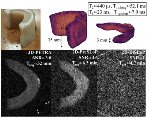
To expand the capabilities of Zero Echo Time (ZTE) pulse sequences with a slice selection method suitable for the shortest-lived tissues in the body. We introduce two new sequences that integrate spin-locking pulses into standard ZTE imaging to achieve slice selection: one for moderately short T2 (DiSLoP), the other for ultra-short T2 samples (PreSLoP). These methods exploit the slower signal decay (at T1ρ≫T2) to retain the magnetization in the slices during the selection process, which is otherwise comparable to or even much longer than T2. We demonstrate control over the slice profiles and positions for 2D imaging. We measure magnetization decay times during spin-locking (T1ρ) as a function of pulse amplitude, showing significant lifetime enhancement for amplitudes as low as 10 uT. We show imaging of slice-selected samples with T2 characteristic times in the range of single milliseconds with DiSLoP and PreSLoP, and with the latter for sub-millisecond T2 tissues. As compared to standard 3D ZTE sequences, PreSLoP achieves the same signal-to-noise ratio (SNR) in 2-5 times shorter scan times, and we argue that this is due to the filling scheme of the finite gap at the center of k-space unavoidable with ZTE sequences. Finally, we discuss a combination of DiSLoP with a dynamical decoupling sequence to avoid this central gap, leading to further scan time accelerations.The proposed sequences are capable of slice-selected 2D imaging of tissues with T2 as low as 275 us with good SNR within clinically acceptable scan times.
Publication: Prepolarized MRI of Hard Tissues and Solid-State Matter.
J. Borreguero, J.M. González, E. Pallás, J.P. Rigla, J.M. Algarín, R. Bosch, F. Galve, D. Grau-Ruiz, R. Pellicer, A. Ríos, J.M. Benlloch, J. Alonso. NMR Biomed. 2022 Apr 5: e4737. doi: http://10.1002/nbm.4737
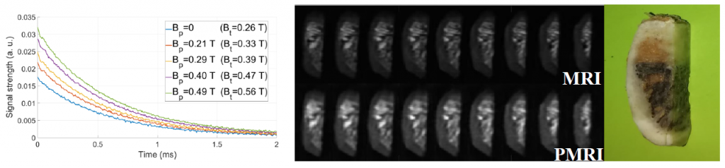
Prepolarized Magnetic Resonance Imaging (PMRI) is along-established technique conceived to counteract the loss in signal strength inherent to low- field MRI systems. When it comes to hard biological tissues and solid-state matter, PMRI is severely restricted by their ultra-short characteristic relaxation times. In the paper, we demonstrate that hard tissue prepolarization is possible with a 0.26T scanner designed for dental MRI. These results can be applied to clinical dental imaging, making low-field PMRI scanners a possible replacement for hazardous X – ray systems.